Actualité internationale
| http://www.lesoir.be/
mercredi 25 août 2010 Des cornées biosynthétiques implantées
chirurgicalement ont permis de restaurer en partie la vue de certains patients.
|
Deux ans après avoir été
implantées, ces cornées restaient totalement fonctionnelles
et ont contribué à la régénérescence,
dans l'implant, de cellules provenant de la cornée du sujet ainsi
que des nerfs sectionnés durant l'intervention, précisent
ces chercheurs dont l'étude paraît dans l'édition du
25 août de la revue médicale Science Translational Medicine,
publiée par le journal Science.
En outre, le réflexe de clignement des yeux et le film lacrymal, fine couche liquide maintenue à la surface de la cornée et protégeant l'épithélium, ont été restaurés chez les participants. L'acuité visuelle s'est améliorée chez six patients, a été inchangée chez deux et a diminué pour deux autres. Aucun n'a subi de réaction de rejet ou de thérapie immunosuppressive, fréquente chez les patients recevant des transplantations d'organe dont des cornées. Les dix patients de l'étude souffraient d'un kératocône avancé, une déformation conique du centre de la cornée progressive et lente. Ils ont eu une implantation d'une cornée biosynthétique dans un seul il. |
| Contact: Vicki Cohn
vcohn@liebertpub.com 914-740-2156 Mary Ann Liebert, Inc./Genetic Engineering News New Rochelle, NY, August 13, 2010
(suite)
|
suite:
Mary Ann Liebert, Inc. (www.liebertpub.com) is a privately held, fully integrated media company known for establishing authoritative peer-reviewed journals in many promising areas of science and biomedical research, including Tissue Engineering, Stem Cells and Development, and Cellular Reprogramming. Its biotechnology trade magazine, Genetic Engineering & Biotechnology News (GEN), was the first in its field and is today the industry's most widely read publication worldwide. A complete list of the firm's 60 journals, books, and newsmagazines is available at our website (www.liebertpub.com). Contact: Vicki Cohn vcohn@liebertpub.com 914-740-2156 Mary Ann Liebert, Inc./Genetic Engineering News Human Gene Therapy http://www.liebertonline.com/1
free access
This review summarizes the current progress and future prospects of recombinant AAV (rAAV)-mediated gene therapy interventions/applications for a wide variety of ophthalmologic disorders. Abstract:
|
| http://www.advancedcell.com/
En collaboration avec le magazine "La Recherche" Cécité, infarctus et même
infertilité, il pourrait bientôt être possible de traiter
de nombreuses maladies à l'aide d'une greffe de cellules. Les premiers
essais sont encourageants.
(suite)
|
suite:
Difficulté qu'a réussi à surmonter, en 2007, l'équipe de Michel Pucéat de l'Inserm à Evry et de Philippe Menasché, chirurgien cardiaque à l'hôpital européen Georges-Pompidou, à Paris : ils ont ainsi pu régénérer des curs de rats victimes d'infarctus ! Pour aboutir à de tels résultats, les chercheurs ont cultivé des cellules souches embryonnaires humaines dans des conditions favorisant leur différenciation en précurseurs de cellules cardiaques. Les cellules obtenues ont été implantées dans le cur de rats victimes d'infarctus. Elles ont alors achevé leur différenciation pour devenir des cellules adultes, capables d'améliorer le fonctionnement du muscle cardiaque. Récemment, les chercheurs ont obtenu des résultats encourageants en greffant des précurseurs de cellules cardiaques, obtenus à partir de cellules souches embryonnaires, à des singes rhésus ayant eu un léger infarctus. De plus, l'équipe de Michel Pucéat et Philippe Ménasché a mis au point une méthode permettant de sélectionner au mieux les cellules cardiaques : ils ont identifié une molécule à la surface de ces cellules, qui permet de s'assurer qu'elles ne sont plus capables de donner naissance à un nombre infini de cellules de tous types. " Cela permet d'éviter toute contamination par des cellules moins différenciées qui pourraient proliférer sans contrôle et former une tumeur ", explique Michel Pucéat. " Nous espérons commencer les premiers essais d'ici deux ans chez des patients ayant eu un infarctus du myocarde de longue date et n'étant pas candidats à une transplantation cardiaque. L'essai pourrait être étendu à d'autres pathologies cardiaques ". Les promesses des cellules souches ne s'arrêtent pas là : elles pourraient venir au secours de l'infertilité. En effet, des spermatozoïdes ont récemment été produits à partir de cellules souches embryonnaires humaines. Une première réalisée par l'équipe du britannique Karim Nayernia de l'université de Newcastle. "Cette découverte permettra de mieux comprendre les causes de l'infertilité masculine en élucidant les mécanismes de différenciation des gamètes, et elle pourra peut-être un jour, être utilisée comme traitement contre celle-ci", s'enthousiasme Samir Hamamah, directeur d'une équipe Inserm, à Montpellier. Ces spermatozoïdes ont été obtenus à partir de cellules ES porteuses des chromosomes X et Y. Une savante manipulation a permis aux cellules souches dotées de 2 lots de chromosomes de n'en avoir plus qu'un, comme tout spermatozoïde qui se respecte. Certains spermatozoïdes avaient un flagelle et pouvaient se mouvoir. Toutefois, " pour être sûr que ce soit de réels spermatozoïdes, il faudrait vérifier leur capacité à féconder un ovule " note Samir Hamamah. C'est ce qu'avait fait trois ans plus tôt la même équipe pour des spermatozoïdes de souris produits in vitro à partir de cellules ES : ce sperme avait donné naissance à des souriceaux. |
| http://www.nature.com/
Elena Ezhkova1 & Elaine Fuchs (1) Abstract Work on stem cells is one of the hottest research areas in biology. But are such studies of any therapeutic value? Fortunately, yes, as is evident from successes in treating blindness. |
Few people today dispute the enormous potential
of stem cells for regenerative medicine. But, despite ever-increasing reports
on the Internet of stem cells being used to treat various disorders from
Alzheimer's disease to spinal-cord injuries to severe heart conditions
proven stem-cell therapies remain few and far between.
1. Elena Ezhkova and Elaine Fuchs are at the Howard Hughes Medical Institute, Rockefeller University, New York, New York 10065, USA. Email: fuchslb@rockefeller.edu |
|
By Courtney Humphries
Scientists have created a three-dimensional,
retina-like structure out of human embryonic stem cells that they hope
could someday serve as a retinal transplant for people with macular degeneration
and other diseases of the retina. Their method, published recently in Journal
of Neuroscience Methods, offers a potential new source of cells for retinal
transplants.
(suite)
|
suite:
Robert Lanza, chief scientific officer at Advanced Cell Technologies, who was not involved in the study, says that his team discovered several years ago that, when turning human embryonic stem cells into RPE cells, other stem cells would spontaneously form layers, including patches of photoreceptors. "This paper shows that you can take advantage of this natural process, and for the first time use tissue engineering techniques to generate three-dimensional retina-like structures," he says. But Lanza is skeptical about the clinical usefulness of such structures. "You can't just transplant a retina and restore sight," he says, because it requires making a series of complex connections with the brain. Although he says there could prove to be some advantage to using three constructs of cells, "for the moment, replacing individual cell types might be the best approach for helping patients suffering from eye disease." Scientists have been working on several approaches to retinal transplants. One approach, led by Advanced Cell Technologies, is to turn human embryonic stem cells into RPE cells and transplant them into the retina. The therapy would work best in the early stages of degeneration to halt further progress, rather than to restore vision that is already lost. Another approach is to transplant stem cells that are in the early stages of becoming light-sensitive photoreceptors, which has demonstrated efficacy in mice. Yet another strategy is to use young tissue instead of individual cells. Fetal tissue transplants have shown some success in animals as well as a small group of humans. A study published in 2008 found that seven out of 10 patients who received the transplants had improved vision. However, there has been debate about whether these transplants actually integrate into the existing tissue. Keirstead has conducted a series of studies in animals that he says demonstrates that transplanted tissue is functioning in the eye. If so, the strategy could be useful for later-stage degeneration, when the existing retina has lost much of its function. For Keirstead's team, the next step is to show that tissue derived from stem cells can function properly. His lab is currently transplanting the tissue into rats to determine whether the transplants can survive and incorporate into the eye, and whether they improve the animals' vision. |
| 19 May 2010 Fraunhofer-Gesellschaft
For many patients who become blind after an accident or illness, a corneal transplantation could restore the ability to see. Each year, 40,000 people in Europe in Germany, about 7,000 await the opportunity to be able to see again, thanks to cornea donors. But donor corneas are not common. Dr. Joachim Storsberg of the Fraunhofer Institute for Applied Polymer Research IAP in Potsdam-Golm developed material and production process for a corneal prosthesis made of plastic. These can help patients who are unable to tolerate donor corneas due to the special circumstances of their disease, or whose donor corneas were likewise destroyed. In recognition of this accomplishment, Dr. Storsberg is being awarded the 2010 Joseph von Fraunhofer Prize. The miniscale artificial cornea has to meet almost contradictory specifications: On the one hand, the material should grow firmly together with the cells of the surrounding tissue; on the other hand, no cells should settle in the optical region of the artificial cornea - i.e., the middle - since this would again severely impair the ability to see. And: The outer side of the implant must be able to moisten with tear fluids, otherwise the implant will cloud up on the anterior side. This would consequently require the patient to get a new prosthesis after a relatively brief period of time. And: The outer side of the implant must be able to moisten with tear fluid, so that the eyelid can slide across it without friction. Dr. Storsberg found the solution with a hydrophobic polymer material. This material has been in use for a long time in ophthalmology, such as for intraocular lenses. In order for it to satisfy the various characteristics required, complex development steps were necessary. The material was thoroughly modified on a polymer-chemical basis, and subsequently re-tested for public approval. |
In order to achieve the desired characteristics, the edge of the implant was first coated with various, special polymers. Then, a special protein was added that contains the specific sequence of a growth factor. The surrounding natural cells detect this growth factor, are stimulated to propagate and populate the surface of the corneal margin. Thus, the cells of the surrounding tissue grow with the implant, and the artificial cornea attains stability. The eye prosthesis evolved jointly with physicians and manufacturers in the EU project, Artificial Cornea." The interdisciplinary research team needed three years to develop the artificial cornea. In a first step, they sent the chemical-biomimetic coated implant to Dr. Karin Kobuch of the Poliklinik für Augenheilkunde at the Regensburg University Medical Center and to the medical center at the Technical University of Munich, on the right banks of the Isar river. The physician examined the artificial corneas in dissected pigs eyes and specialized cell cultures. Eventually, the team under Dr. Gernot Duncker and Dr. Saadettin Sel of the University Center for Ophthalmology in Halle (Saale) tested the more complex models in rabbits. There, the design was further refined: the optics were made smaller, and the implant haptic enlarged in order to maintain a more stable construction. Miro GmbH manufactured the implant, robin GmbH handled the distribution and sales and supported the specially adapted implantation centers in Europe. By 2009, a prosthesis was already successfully in use; further implantations are anticipated during the first six months of 2010. |
| Web-connected cameras may help doctors
detect a common eye disease.
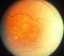
By Arlene Weintraub Friday, May 14, 2010 Of all the complications of diabetes, few are as devastating as diabetic retinopathy, a progressive eye disease that causes blurred vision and in some patients, blindness. By the time most patients recognize something's wrong, it's often too late for them to be treated effectively. As a result, diabetes is the leading cause of vision loss among adults over 20. More than 12,000 new cases of blindness each year are caused by diabetic retinopathy, according to the National Institutes of Health. 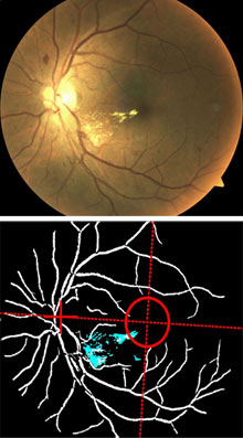
Eye sight: White dots a patient's retina (top) are early signs of disease. In the image below these spots are identified automatically. Credit: Ken Tobin An ophthalmologist and a scientist from the
Department of Energy's Oak Ridge National Laboratories in Tennessee believe
they can help doctors detect diabetic retinopathy long before the disease
wreaks havoc on their patients' vision. Their startup company, Automated
Medical Diagnostics (AMDx), has developed software that can detect the
early signs of diabetic retinopathy by comparing digital photos of a patient's
retina to images that represent various stages of diabetic eye disease.
AMDx's founders believe their technology will enable all health workers--even
those who are not trained in eye care--to take retinal scans of any patient,
zap them over the Internet to AMDx's servers, and get a diagnosis back
before the patient leaves the office. "We're trying to show we can be
as accurate as a trained ophthalmologist," says Ken Tobin, AMDx co-founder
and division director of measurement science and systems engineering at
Oak Ridge.
(suite)
|
suite:
(...) That makes it easier for the technology to detect lesions, leaky blood vessels, swelling, and other abnormalities on the retina that can be early signs of disease. Chaum and Tobin spent five years developing algorithms that can extract information from retinal images and screen it against a database of more than 20,000 photos. AMDx doesn't produce diagnoses but rather alerts doctors to patients who need to be referred to specialists for more in-depth testing, diagnoses and treatments. AMDx is currently testing its system in a handful of clinics in Mississippi and Tennessee. Training doctors is easy, Chaum says, because the cameras have features like auto-focus, and they don't require that the patients' eyes be dilated. The photos are sent over the Internet to AMDx's servers and automatically compared to images in its database. Chaum then checks each result manually--a process that takes about 90 seconds per case, he says. AMDx's goal is to ultimately turn over the entire job to its computers, but for now it must rely on Chaum's review. That's because some insurers--most notably Medicare and Medicaid--will only reimburse physicians for eye screenings after an ophthalmologist examines the results. Chaum and Tobin are collecting data, with the goal of proving to both regulators and insurers that their computers are as effective as Chaum at detecting disease. "The computers can handle thousands of reports a day. The bottleneck is me signing off on them," Chaum admits. Efforts to remotely diagnose eye diseases have been tried on a limited basis by the Veterans Administration hospitals and other institutions. Some ophthalmologists believe that if the idea catches on, they'll be able to treat many more cases of diabetic retinopathy than they can today. "If you can teach a clinician to recognize changes in the eye, you can teach a computer to do it," says Barrett Katz, an ophthalmologist and professor at Montefiore Medical Center and the Albert Einstein College of Medicine in the Bronx, NY. "It doesn't take an ophthalmologist to gather these images--if anyone could do it, that would be a major step forward." Tobin and Chaum are on the hunt for venture capital to fund AMDx's expansion. They hope the current focus on health reform will give them a boost, because the ongoing debate is drawing attention to the need for improving the efficiency of the health care system. "It's not that we can't treat diabetic retinopathy, Chaum says. It's that we're inefficient in how we screen for it." Related Articles
 » A Stem-Cell Therapy for Blindness: 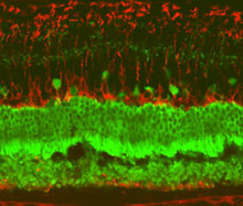 » Eye Implants to Fight Progressive Blindness:  |
| 17 May 2010 Durham University
Thousands of people who are
partially-sighted following stroke or brain injury could gain greater independence
from a simple, cheap and accessible training course which could eventually
be delivered from their mobile phones or hand-held games consoles, according
to a new study.
(suite)
|
suite:
The scientists say patients may even be able to see similar improvements in their vision by playing mainstream computer games, particularly those whereby you need to scan virtual environments with your eyes. Professor David Mendelow, a neurosurgeon at Newcastle General Hospital and professor of neurosurgery at Newcastle University, said: "Hemianopia is often not recognised and is probably much more common than realised. Patients and their families find it very difficult to understand this problem of "half blindness". "At Newcastle General, we have trained our occupational therapists to recognise this visual problem and we can now identify patients with hemianopia at an early stage. "The Neurosciences Unit at Durham University, where we refer patients on to, is to be congratulated on demonstrating how successful this kind of visual retraining can be." CASE STUDY Nichola Burlison from Low Willington, County Durham*
* Please note that we are not able to give any further biographical details due to the nature of the case study's employment. FACTS ABOUT HEMIANOPIA
http://www.durham.ac.uk/news
|
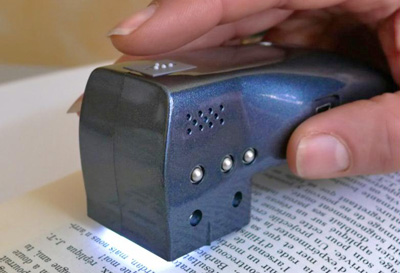
| AFP
A partir de tout document écrit, un livre, une revue ou une notice de médicaments, le boîtier à lecture optique retranscrit un texte en braille pour les aveugles et mal voyants, grâce à des picots qui surgissent au fur et à mesure de la lecture. Il est également doté d'une fonction audio pour transformer le texte en son. "Nous allons pouvoir dire aux aveugles et mal voyants qu'ils ont un produit qui peut leur simplifier la vie", a déclaré le lauréat, ancien professeur de mathématique et ingénieur de formation, qui espère voir le prix de vente (1.680 €) de son invention baisser considérablement pour le rendre accessible au plus grand nombre. "Nous n'avons vendu que 150 appareils (via sa société Vision SAS), c'est très peu au regard des 100.000 mal voyants et aux 250.000 très mal voyants en France", a-t-il encore souligné. |
Autre invention primée: le Top-reader
"Je pensais à cet appareil depuis de nombreuses années parce que ma soeur malvoyante n'avait de cesse de demander l'utilité d'apprendre le braille alors que très peu de documents sont écrits en braille", a raconté Raoul Parienti, peu avant la remise des prix. Ce sont dix années de recherche et développement et 1,5 million € qui ont été investis dans ce projet. L'appareil lit actuellement des textes en sept langues: français, anglais, espagnol, italien, allemand, portugais et néerlandais, car le premier donateur est hollandais. Mais son concepteur aimerait bien le décliner dans toutes les langues, pourquoi pas en hiéroglyphes ou en chinois. Outre le premier prix, Raoul Parienti, qui a déposé par le passé plus de 100 brevets d'innovation, a remporté le troisième prix pour son "Top-reader", le pendant du "Top-braille", un appareil permettant cette fois la lecture de tout texte photographié et numérisé. Plus de 500 inventions ont été présentées lors de cette 109e édition du célèbre concours qui a vu naître le stylo à bille, le fer à repasser à vapeur, le four électrique ou encore le coeur artificiel. |